
Reviewed personally by Dr. Sunkara · Written for Chronic Subdural Haematoma Treatment in Hyderabad · Updated October 2026
Quick Answer
Chronic subdural haematoma treatment in Hyderabad addresses a slow collection of old blood, known as a chronic subdural haematoma or SDH, between the brain's surface and its outer covering. It usually follows a minor fall weeks earlier, often in older adults or people on blood thinners. Small, stable collections are sometimes watched; surgery, usually burr-hole drainage, treats the rest.
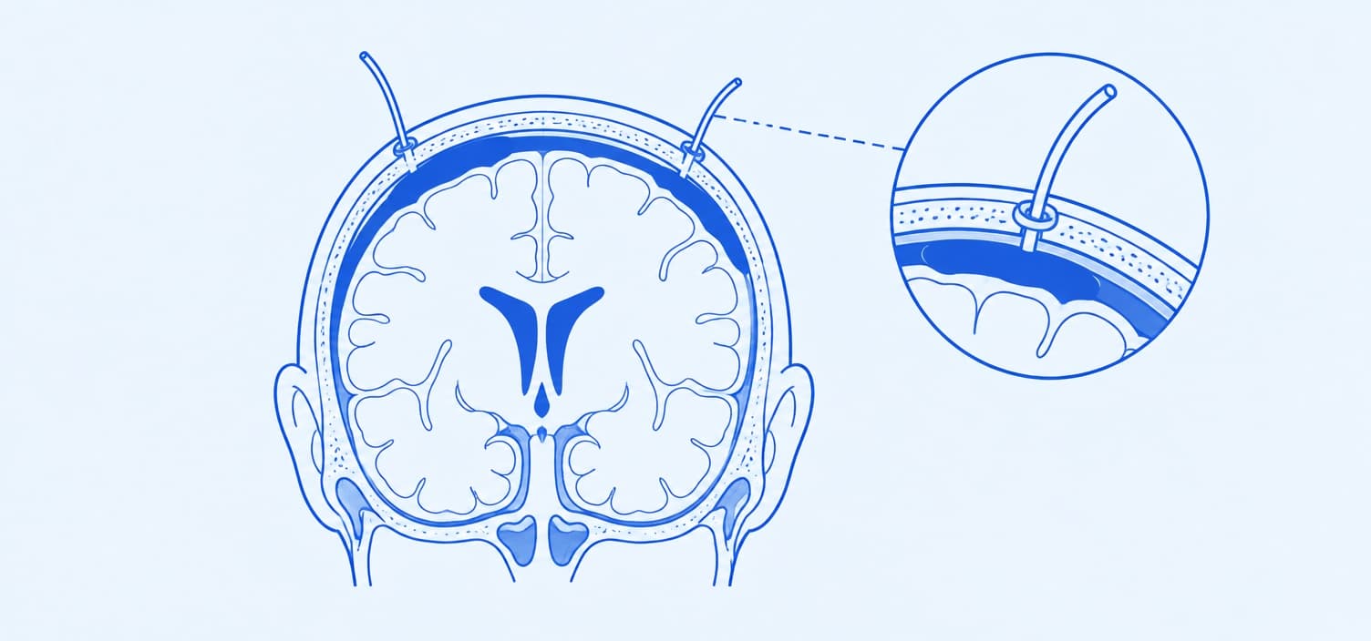
Most chronic subdural haematomas trace back to a fall that felt minor at the time, sometimes months before symptoms show up. Head injury treatment covers that early stage. Head Injury Treatment → What happens when the bleed keeps growing quietly for weeks is the part most people never hear about.
One recent patient, who had mild Parkinson's on medication, assumed his growing weakness was simply his Parkinson's progressing. A routine follow-up scan instead found a chronic SDH, and his symptoms improved drastically after surgery. Not every chronic SDH needs an operation though: small, stable ones are often just watched. Surgery enters the picture once the haematoma presses on the brain or symptoms build, and it's rarely as invasive as patients expect.
Chronic SDH treatment in Hyderabad becomes relevant once a slow bleed starts affecting how someone thinks, moves or feels.
01
A CT or MRI scan confirms the haematoma's size and location, and Dr. Sunkara weighs whether it needs draining now or can be safely watched.
02
One or two small burr holes are drilled into the skull under anesthesia, and the old, liquefied blood is drained out through a soft tube.
03
A drain often stays in place for a day or two, and a follow-up scan checks that the brain has re-expanded and the space hasn't refilled.
What does getting ready for chronic SDH surgery involve? It's less daunting than it sounds.
01
The standard approach for most chronic SDH cases: one or two small holes let the old blood drain out, often with a tube left in place to catch any that reaccumulates.
02
A smaller, single-hole technique done under local anesthesia at the bedside in select cases. It can suit frail, elderly patients who may not tolerate a longer procedure well.
03
A small bone flap is opened for haematomas with thick membranes or solid clot that a burr hole alone can't adequately drain, giving more direct access.
04
A newer, minimally invasive option that blocks the small artery feeding the haematoma's outer membrane, sometimes used alongside surgery or for haematomas likely to recur. Dr. Sunkara's interventional radiology colleague performs this.
01
Close monitoring on the ward or in a step-down unit, watching for new weakness, confusion or a change in alertness as the brain re-expands.
02
2 to 3 days before discharge, once the drain is out and a follow-up scan looks stable.
03
Most patients return to light daily activity, with energy and clarity of thought improving gradually as the brain settles.
04
A follow-up CT or MRI confirms the haematoma hasn't reaccumulated and checks that the brain has fully re-expanded to fill the space it left.
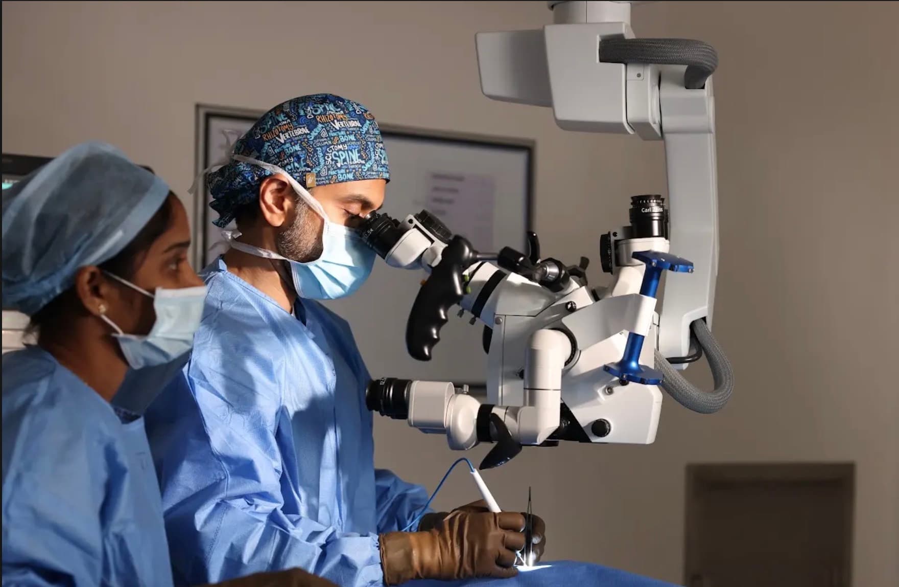
Most chronic SDH patients improve significantly once the pressure on the brain is relieved.
Chronic subdural haematoma treatment in Hyderabad carries real risks, like any procedure, but serious complications are less common than most patients expect.
What The Risks Include
It's a slow buildup of old blood between the brain's surface and its outer covering, usually after a minor head injury. Symptoms often take weeks to appear.
Chronic subdural haematoma treatment in Hyderabad starts with a clear read of your scan, whether it's a new finding, a recurrence, or a second opinion.

Written & reviewed by Dr. Raveesh Sunkara, Senior Consultant Neurosurgeon & Spine Surgeon · Last updated October 2026 · Next review due April 2027